TEAM NET FENG
Priority 2 of the Programme: European Funds for a Modern Economy 2021–2027 (FENG)
Grant Agreement No. FENG.02.03-IP.05-0113/24
Project value (total project cost): PLN 12,344,749.02
Amount of European Funds contribution: PLN 12,344,749.02
Development of an innovative, high-throughput platform for functional screening of human pancreatic endocrine cells
About Project
Diabetes mellitus, a metabolic disorder with increasing global prevalence, arises from an imbalance in glucose metabolism. This imbalance is largely attributed to defective pancreatic beta cells, the primary producers of the hormone insulin, residing within the islets of Langerhans. Dysfunction of these beta cells leads to hyperglycemia, a condition characterized by abnormally high blood glucose concentrations.
To expand therapeutic options for individuals with diabetes efficiently, a source of human pancreatic beta cells is crucial. Currently, the most promising avenue involves the in vitro differentiation of pluripotent stem cells into insulin-producing and secreting beta cells. Recent research from multiple laboratories worldwide, including our own, has demonstrated the potential to generate human beta cells that, in many aspects, resemble their native counterparts.
In this study, we propose to explore the characteristics of these newly generated beta cells to improve both their quality and quantity. Furthermore, recognizing the intensive crosstalk between beta cells and other cell types within the pancreas, specifically endothelial cells, we also propose investigating the molecular nature of this communication using genomic screening, microfluidics, and in vivo diabetes models. Understanding these interactions is vital for further enhancing the accuracy and function of in vitro differentiated human beta cells. Such pancreatic organoids will offer a unique opportunity for precision in drug discovery.
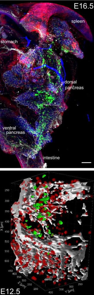
(lower panel) 3D model of the developing pancreas and its microenvironment in a 12.5-day-old mouse embryo. Green spots represent beta cells, while the white and red areas indicate blood vessels and mesenchyme.
Project Team
- Uniwersytet im. Adama Mickiewicza w Poznaniu – prof. UAM dr hab. Małgorzata Borowiak – Instytut Biologii Molekularnej i Biotechnologii – Zakład Ekspresji Genów – https://ibmib.web.amu.edu.pl/groups/stem-cell-laboratory/
- Uniwersytet Warszawski – dr inż. Tomasz Kamiński – Instytut Biochemii -Zakład Biologii Molekularnej –https://www.biol.uw.edu.pl/jednostki-naukowo-dydaktyczne/instytut-biochemii/zaklad-biologii-molekularnej/
- Uniwersytet Jagielloński -Prof. dr hab. Józef Dulak -Wydział Biochemii, Biofizyki i Biotechnologii UJ -Zakład Biotechnologii Medycznej –https://zbm.wbbib.uj.edu.pl/kontakt1
News
- Competition for the position of a senior post-doc / bioinformatician researcher at the Faculty of Biology AMU
- ADAM MICKIEWICZ UNIVERSITY, POZNAN ANNOUNCES A COMPETITION for:
post-doctoral researcher position
specialist position at the Faculty of Biology in the TEAM NET project:
post-doctoral researcher position at the Faculty of Biology in the TEAM NET project:
- Job offers
- 2 Postdoctoral Positions in the Project
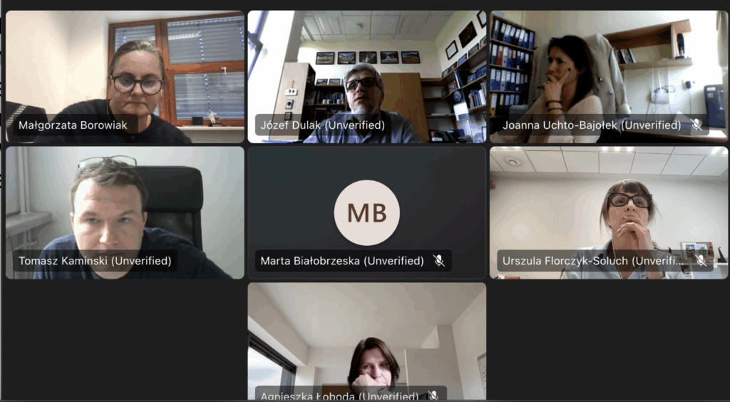
- TEAM-NET Kick-off: Collaboration and Innovation in Action (April 24, 2025)
Our recent kick-off meeting brought together members from all three participating teams. It was a productive session filled with in-depth planning and the emergence of exciting ideas that will drive our project forward.
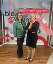
- Bioforum insights: bridging the gap between research and commercialization (April 2-3, 2025, Warsaw)
Our project’s Broker, Alicja Ostrowska-Leszczynska, and the UAM team leader, Prof. Malgorzata Borowiak, represented TEAM-NET at the Bioforum meeting in Warsaw (April 2-3, 2025). Their participation focused on engaging with representatives from start-up companies and the pharmaceutical industry to discuss the key challenges associated with translating significant research findings into viable commercial products.
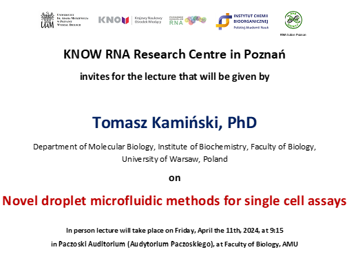
- Microfluidic innovations at IBMiB Poznan (April 11, 2025)
Dr. Tomasz Kaminski, our UW team leader, delivered an inspiring talk on “Novel droplet microfluidic methods for single cell assays” at IBMiB in Poznan. The talk was well-received, fostering inspiration, showcasing cutting-edge technology, and sparking exciting discussions around this innovative technology.
- TEAM-NET Project officially launched! (April 1, 2025)
We’re thrilled to announce the official launch of our TEAM-NET project, proudly funded by the Polish Foundation for Science (FNP). This exciting endeavor wouldn’t have been possible without the exceptional support of our administrative coordinators: Iwona Kanonik-Jedrzejak (UAM), Katarzyna Sienkiewicz (UW), and Joanna Uchto-Bajolek (UJ). A huge thank you to them for their invaluable contributions!